Microscopy and Photography: Capturing the Unseen
Written on
Chapter 1: The Art of Visualization
Microscopy serves as a powerful tool for examining the intricate details of tissues and cells, allowing scientists to visualize the presence, location, and behavior of various cellular components. In many ways, microscopy parallels photography; both require a clear understanding of what you wish to capture and how to achieve that vision.
A conversation with my supervisor about epifluorescent microscopy highlighted this analogy. He cautioned me against bleaching my cells with fluorescent light, likening the practice to photography. Initially, I disagreed, but later I recognized the truth in his statement.
In photography, capturing an unexpectedly beautiful scene—like a breathtaking sunrise on a frigid winter morning—can lead to a completely different interpretation when repurposed in another context, such as a blog post on Medium.
Returning to microscopy, the goal is to discern the inner workings of cells to understand their components better. If you lack clarity on what you intend to see or how to observe it, your efforts may not yield the desired insights.
Section 1.1: Defining Your Focus
As a researcher studying airway mucus, I investigate whether specific treatments affect mucus secretion levels. To differentiate cuboidal epithelium in the Air Liquid Interface Culture system, I employ staining techniques using the MUC5AC vesicle marker alongside the Beta Tubulin marker for ciliated cells.
At this point, we can explore two approaches—live cell imaging or examining fixed cells. For the sake of simplicity, we'll focus on fixed cells. To visualize various cellular depths, I choose epifluorescent microscopy, which provides comprehensive depth information.
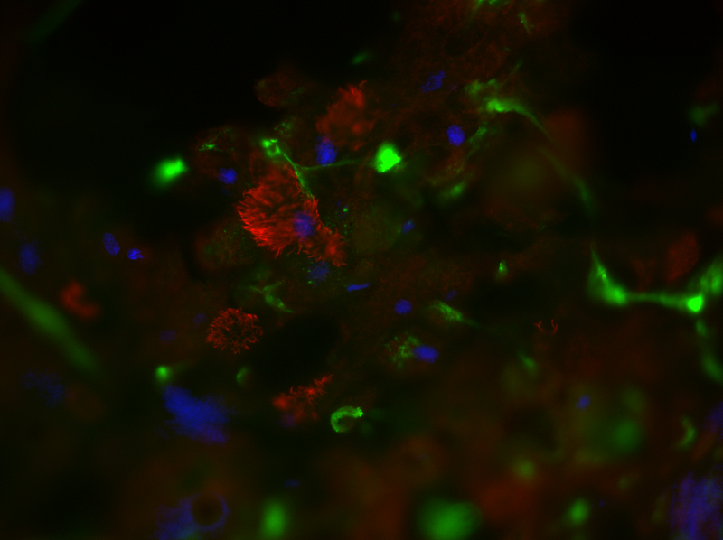
Figure 2. Epifluorescent microscopy image showcasing ciliated and goblet cells, with distinct colors representing various cellular components.
Utilizing Z-stacking, I can obtain layered images from the apical surface to the basal layer. This technique allows for video recordings traversing through the layers or capturing still images. However, it’s essential to note that neither brightfield nor confocal microscopy can be entirely substituted for this method.
Section 1.2: Exploring Confocal Microscopy
Confocal microscopy offers an even more refined approach to visualization. Imagine shining a flashlight into a dark tunnel; this is akin to how confocal microscopy operates. By directing laser light precisely within a tissue or cell layer, you can focus on a specific plane while minimizing blur from other areas.
Like Z-stacking, confocal microscopy requires the same method of layering, but it selectively captures sharp images from particular depths, allowing for detailed analysis. The ability to focus on a specific transverse section of a plane opens intriguing possibilities for medical diagnostics.
Chapter 2: The Importance of Knowledge
To effectively capture images using microscopy, a solid foundation in basic biology is crucial. Lacking this knowledge may result in images that appear abstract or nonsensical—merely indistinct blobs of cells. Understanding how to focus correctly, especially for those with visual aids, is vital for achieving high-quality images.
When quantifying specific cell types, like FTO knockout cardiomyocytes, familiarity with the structures, fluorophore spectra, and size scales is necessary. Misidentifying a nucleus could lead to significant errors in interpretation.
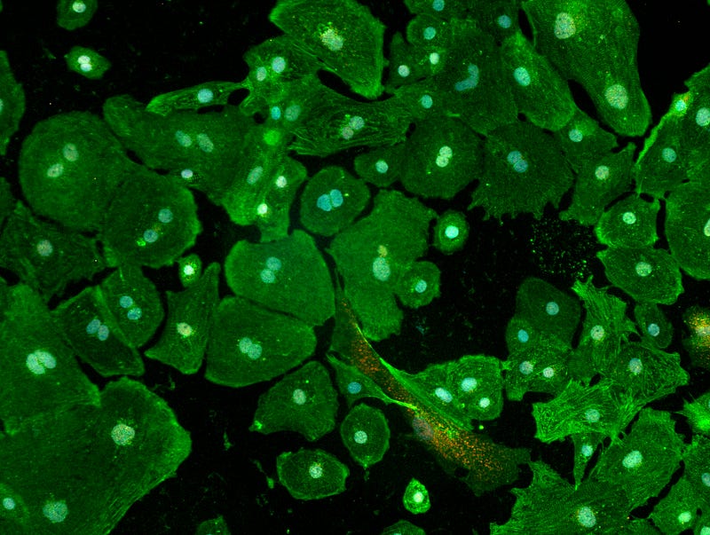
Figure 3. Fluorescent microscopy image illustrating cardiomyocytes with unclear cell borders, typical for animal cells.
It's important to recognize that unlike plant cells, animal cells lack clear boundaries. With brightfield microscopy, this distinction becomes apparent. Additionally, understanding potential contaminants—like plastic artifacts or lab coat fibers—is essential for accurate observations.
Section 2.1: Ethical Considerations in Image Processing
While processing images using software like Fiji ImageJ is common, one must tread carefully. Assigning colors to separate channels and merging them to produce aesthetically pleasing images must be balanced with ethical considerations.
Although minor adjustments in brightness and contrast are generally acceptable, significant alterations that misrepresent data are not. Transparency regarding raw data is crucial, as most journals require it for validation.
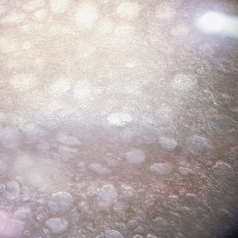
Figure 4. An image depicting contamination in cell culture, which can affect experimental results.
As a researcher, consulting with a supervisor regarding image processing techniques can provide valuable guidance. Proper justification for any modifications is essential, as excessive alterations may lead to serious repercussions.
The first video, "Microscope Club Taking Photographs," showcases the art and techniques of microscopy in capturing cellular images, emphasizing the parallels with photography.
The second video, "How do you use a Light Microscope? A step-by-step guide!" provides a detailed overview of light microscopy techniques, essential for effective imaging.
DISCLAIMER:
The images presented here are intended for educational purposes, taken by me, and anonymized from specific experimental conditions. Happy micrography! ;)